Engineering
 Advanced experience in personalized implant design, in-depth understanding of human anatomy and close cooperation with practicing surgeons – those are the key factors that allow us to overcome the most difficult challenges that can arise during the design and manufacturing phases of creating a personalized implant. Our bioengineers form a foundation of accumulated knowledge via attendance during implant installation, analysis of the surgery’s course and patient rehabilitation as well as attention to surgeon feedback and suggestions. This lets our specialists use the entire base of accumulated knowledge and experience to achieve perfect results.
The specialists of ITC Endoprint regularly exchange experience with European colleagues during international conferences and participate in the world’s largest medical conventions and exhibitions dedicated to implantology and the latest tendencies in additive manufacturing. This way we are able to expand the application of additive manufacturing in medicine and discover unique and revolutionary solutions that advance the sector of endoprosthesis and implantology.
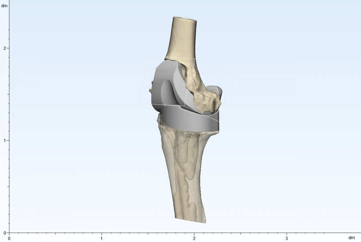
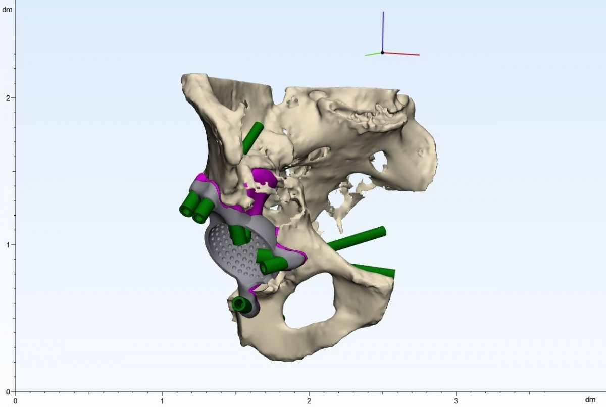
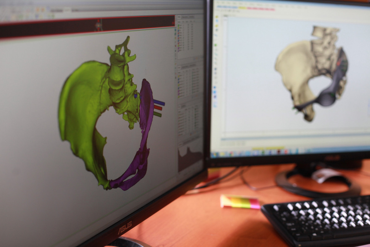
Our company employs the most cutting-edge specialized 3D software that allows our bioengineers to create virtual models reflecting the patient’s anatomy. The patient’s CT/MRI scan files are converted into a 3D model that closely mirror the preoperative condition of the affected area, which is vital for revision surgery planning. Previously installed implants are often prone to produce visual noise in the CT/MRI scan, making the scan difficult to decipher for the attending physician without additional processing. The software used by our bioengineers enables us to eliminate visual noise and separate existing implants, screws and bone cement (commonly used as an anchor for endoprosthetics) from bone structures as well as indentify potentially hidden internal defects. After a 3D model is processed, it then undergoes a process of specialized anatomical analysis, during which anatomical axes and symmetry are restored, thus ensuring correct rotation and complete fuction of all joints related to the affected area by the end of the postoperative period.
A 3D model created this way serves as a basis that allows the attending surgeon and the bioengineer to establish a surgery plan and design auxiliary instruments required for preoperative planning and performing the surgery.
Design stages

DICOM file
(CT/MRI)

3D model
creation

Design in cooperation
with the doctor

Approval

Manufacturing